Gynecomastia diagnosis in Korea: why high-resolution ultrasound matters more than self-diagnosis
Patient Registration Info
| Age | Weight | Height | BMI |
|---|---|---|---|
|
30s |
71kg |
172cm |
24 |
Surgical site
Surgical area: Gynecomastia | Grade 2-A
Can every firm lump in the chest be considered gynecomastia?
Not every firm lump felt inside the chest
is diagnosed as the same type of gynecomastia.
Because both fat and glandular tissue
may be present in the male chest,
it is difficult to determine the cause
based on touch alone.
An ultrasound examination is needed to evaluate
the development of glandular tissue
and the distribution of surrounding fat.
[Learn More About Lesarts Gynecomastia Surgery]
Many patients considering gynecomastia surgery
check their chest for firm lumps before visiting
the clinic or search online for ways
to self-diagnose gynecomastia.
When a firm area is felt inside the chest,
it is often assumed to indicate
the presence of developed glandular tissue.
On the other hand, if the chest feels soft,
some may assume that the fullness
is simply caused by excess body fat.
However, male gynecomastia often presents
as a mixed type involving both
glandular tissue and excess fat.
The perceived firmness of a lump can also vary
from person to person, making it difficult
to accurately determine the condition
through self-examination alone.
Therefore, if gynecomastia is suspected,
it is important not to rely solely
on chest size or how the tissue feels.
A high-resolution ultrasound examination can evaluate
the thickness and distribution of glandular tissue
as well as the amount of surrounding fat.
The patient featured in this case
visited our clinic after becoming concerned
about chest protrusion in his 30s.
He had experienced little recent weight change
and regularly participated in weight training
and played soccer.
However, even as his body fat decreased,
there was little noticeable change
in his overall chest size or
the central protrusion of his chest.
As the chest contour remained unchanged
despite regular exercise, he began to suspect
that the problem might not be caused
by excess fat alone and decided
to undergo a gynecomastia evaluation.
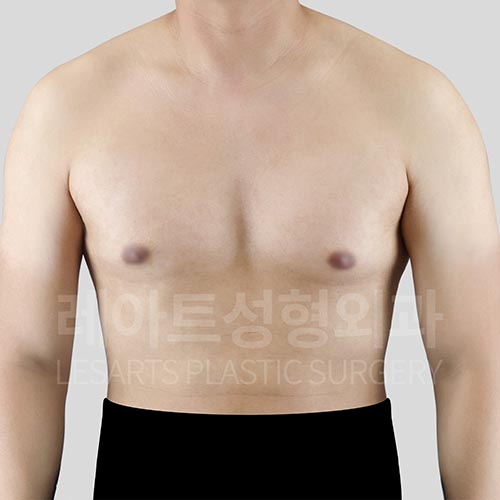
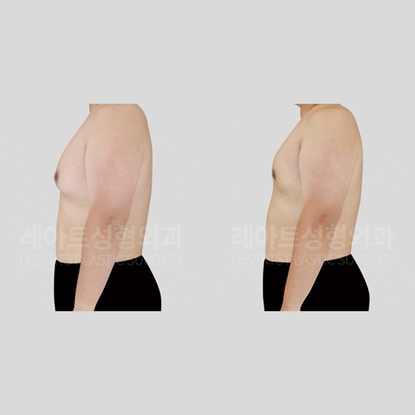
[Preoperative Chest Size and Shape Before Gynecomastia Surgery at Lesarts Plastic Surgery]
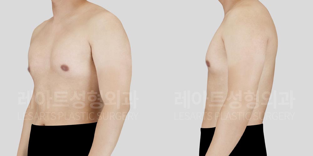
[Gynecomastia Since His 30s: Surgical Plan to Address Both Glandular Tissue and Fat]
[Patient Information and Gynecomastia Evaluation]
Age: 30s
Height / Weight: 172 cm / 71 kg
Diagnosis: Simon Grade 2A, true gynecomastia
Skin Elasticity: Slightly reduced
Before surgery, the central areas of both breasts
projected forward, with additional fullness extending
toward the lower chest and underarm areas.
On palpation, firm lumps could be felt
inside the chest, and high-resolution ultrasound
also confirmed broadly developed glandular tissue
on both sides.
The external appearance corresponded to Simon Grade 2A.
Fat was also present in the lower and outer chest,
making glandular tissue removal alone insufficient
to create a smooth overall chest contour.
We therefore planned gynecomastia surgery to remove
the developed glandular tissue while performing liposuction
of the surrounding fat to evenly reduce volume
in the central and lower chest.
The patient had previously undergone nipple reduction surgery
but remained concerned about the size of his nipples
even after the procedure.
Performing gynecomastia surgery and nipple reduction surgery
at the same time could place additional stress
on the skin and blood supply
in the previously operated area.
For this reason, we decided to perform
the gynecomastia surgery first and reconsider
additional nipple reduction after allowing
sufficient time for recovery.
▶ View More Gynecomastia Surgery Cases
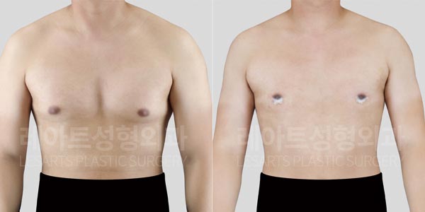
In the following photos taken 2 weeks
after gynecomastia surgery, we will examine changes
in chest protrusion from the front and side,
as well as the chest contour under clothing.
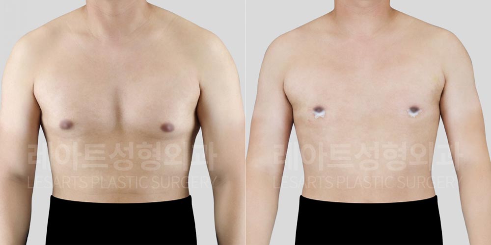
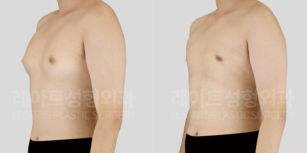
[2 Weeks After Male Gynecomastia Surgery: Changes in Bilateral Chest Protrusion from the Front View]
These photos were taken 2 weeks
after gynecomastia surgery.
Before surgery, the volume of both breasts
created a rounded contour, making the entire chest
appear wider and heavier.
After surgery, the volume in the central
and lower chest was reduced, bringing both sides
closer to the natural contour of the rib cage.
Although some swelling and tissue firmness
remain at this early stage of recovery,
the bilateral chest protrusion is noticeably reduced,
creating a lighter and more balanced upper-body contour.
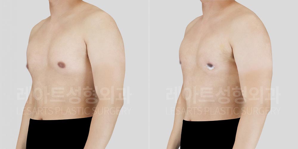
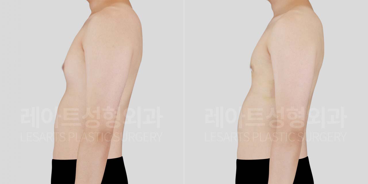
[2 Weeks After Gynecomastia Surgery: Changes in Chest Protrusion and Lower Chest Contour from the Side View]
[2 Weeks After Gynecomastia Surgery: Changes in Chest Protrusion and Lower Chest Contour from the Side View]
From the side view, the forward projection
of the chest has decreased, creating
a flatter transition from the rib cage
to the chest.
Before surgery, volume in the central
and lower chest created visible shadowing
beneath the chest and made the transition
to the upper abdomen appear heavier.
After surgery, the remaining fat layer
in the lower chest was also reduced,
decreasing the shadow beneath the chest
and creating a smoother transition
between the chest and upper abdomen.
Because both the glandular tissue and surrounding fat
were addressed together, the central chest
does not appear abruptly hollow and transitions
more evenly into the surrounding chest contour.
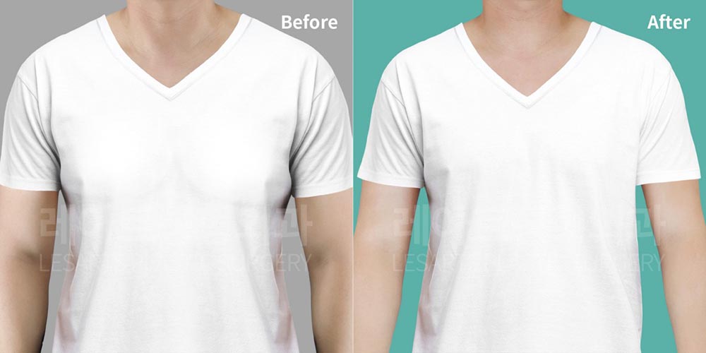
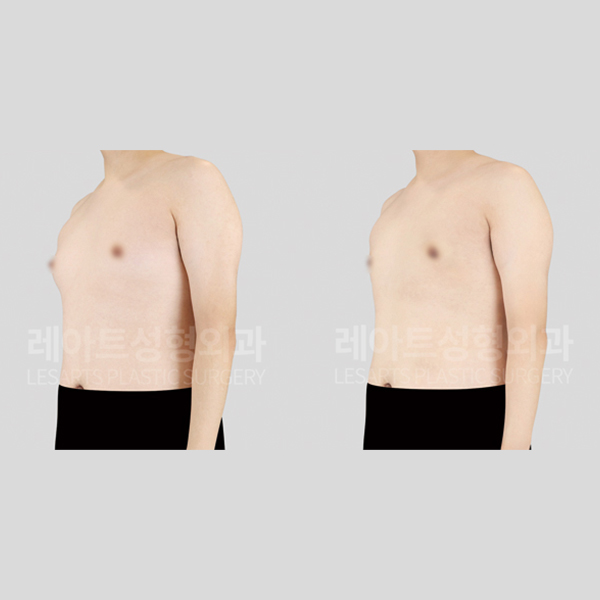
[2 Weeks After Gynecomastia Surgery: Changes in Chest Protrusion and T-Shirt Fit]

[2 Weeks After Gynecomastia Surgery: Side-View Changes in Chest Protrusion and T-Shirt Fit]
The changes in the chest contour
are also clearly visible when wearing clothing.
Before surgery, when wearing a thin T-shirt,
the central and lower chest volume showed through
the fabric, making the patient self-conscious
when choosing tops.
After surgery, the thickness of the chest decreased,
allowing the T-shirt to fall more naturally
along the upper body without catching
on the chest area.
The patient had exercised consistently for years,
but the shape of his chest remained unchanged.
After surgery, he was especially satisfied
that wearing thin T-shirts and workout clothes
felt more comfortable.
When symptoms of gynecomastia appear, some patients
rely on online information and self-diagnosis
or try to conceal their chest
with thicker clothing or posture.
However, if the chest suddenly becomes larger,
a firm lump can be felt, or pain develops,
it is important to accurately evaluate
the tissues that make up the chest.
If chest protrusion remains despite regular exercise
and weight loss, the possibility of developed
glandular tissue should also be considered,
rather than assuming fat is the only cause.
The treatment plan for gynecomastia is not determined
by the visible size of the chest alone.
The thickness and distribution of glandular tissue,
surrounding fat, skin elasticity, and previous surgical history
should all be evaluated before determining
the appropriate treatment range.
#gynecomastiadiagnosis
#gynecomastiaultrasound
#malebreastreduction
#lesartsplasticsurgery
Gynecomastia Surgery FAQ
Q1. Does a firm lump in the chest always mean true gynecomastia?
A firm area may suggest the presence of developed
glandular tissue, but touch alone cannot provide
an accurate diagnosis.
An ultrasound examination is needed to evaluate
the thickness and distribution of glandular tissue.
Q2. If my chest feels soft, is it just excess fat?
Even if the chest feels soft, glandular tissue
may still be present beneath the fatty tissue.
Developed glandular tissue cannot be removed through exercise alone.
If central chest protrusion remains despite consistent exercise,
an examination may be recommended.
Q3. Why is an ultrasound examination necessary for gynecomastia?
Ultrasound can evaluate the extent and thickness
of glandular tissue that may be difficult
to assess through visual examination alone.
It also shows the distribution of surrounding fat,
helping determine whether gland removal, liposuction,
or a combination of both may be needed.
Q4. Why does chest protrusion remain even after exercise?
Body fat may decrease through exercise and weight loss,
but developed glandular tissue cannot be removed
through exercise alone.
If central chest protrusion remains despite consistent exercise,
it may be helpful to undergo a gynecomastia evaluation.
Q5. Can gynecomastia surgery and nipple reduction be performed together?
This depends on the condition of the skin
and any previous surgical history.
If there is concern about skin damage
or reduced blood supply, the procedures may be staged,
with the second surgery planned after sufficient recovery.
Rather than relying solely on self-diagnosis,
a high-resolution ultrasound examination can help determine
the current condition and appropriate treatment options.
▼ Visit Lesarts Plastic Surgery in Gangnam, Seoul, Korea ▼