Simon Grade 2A, True Gynecomastia Surgery 2-Month Postoperative Review
Information
- Age : mid 30’s
- Height : 170cm
- Weight : 79kg
- BMI : 28
Surgical site
- surgical site : gynecomastia
- grade : 2-A
When men notice an increase in chest size and protrusion, they often assume it is simply due to fat accumulation from weight gain.
However, if the chest size and the puffiness around the areolas do not improve despite diet and exercise, it may be a sign of true gynecomastia (glandular hypertrophy).
True gynecomastia is not just a body shape issue but is often caused by hormonal imbalances. If it causes discomfort or concerns, it is essential to seek a medical consultation as a priority.
Even with weight loss efforts, reducing chest size is challenging, and chest exercises alone cannot change its shape. Many men struggle with psychological distress and a lack of confidence due to this condition.
Additionally, some individuals experience gynecomastia symptoms from puberty, leading to long-term stress. When the condition persists regardless of weight changes, it can even negatively impact personality development. Therefore, having accurate information and a proper understanding of gynecomastia surgery is crucial.
In this article, we will discuss a real case of gynecomastia that could not be resolved through chest exercises or dieting. By removing glandular tissue and performing liposuction, the issue was fundamentally corrected. We will explain the diagnostic process, surgical procedure, and post-surgical changes in detail.

[Lesarts plastic surgery before gynecomastia surgery]
This patient is a man in his mid-30s who has been experiencing gynecomastia symptoms since adolescence. During his teenage years, he was close to being overweight and noticed an increase in chest size and protrusion of the areolas.
Even after losing over 10 kg as an adult, the protruding chest and enlarged areolas did not improve at all.
These symptoms caused significant discomfort in daily life, leading him to seek a permanent solution through gynecomastia surgery at our clinic.

[Lesarts plastic surgery before gynecomastia surgery]
The shape of the patient’s chest was visibly protruding.
To determine the cause of the large and protruding chest, a gynecomastia ultrasound examination was performed. The results showed fibrous glandular tissue widely distributed in the areolar area.
The patient was diagnosed with true gynecomastia, classified as Simon Grade 2A.
The medical team thoroughly explained to the patient that simple chest exercises, dieting, or liposuction alone would not be sufficient to improve the protrusion of the chest. Therefore, the plan for gynecomastia surgery to fundamentally reduce the chest size was outlined as follows:
- The glandular tissue would be carefully removed to directly address the cause of the large and protruding chest.
- Excess fat in the lower and lateral chest areas, which contributes to the volume, would be removed through liposuction, ensuring the overall chest contour becomes smooth and natural.
- Post-surgery, measures would be taken to prevent the nipple and areola area tissues from adhering to the underlying fascia, ensuring that no inversion or chest shape imbalance occurs. The tissue would be repositioned to maintain a balanced appearance.
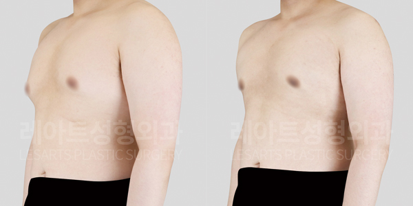
Below are the pre-surgery and two months post-surgery comparison photos of the patient.

[Lesarts plastic surgery before&after gynecomastia surgery(Taken under the same conditions 2 months after gynecomastia surgery.)]

[Lesarts plastic surgery before&after gynecomastia surgery(Taken under the same conditions 2 months after gynecomastia surgery.)]
In the pre-surgery photo of gynecomastia, both sides of the chest show prominent protrusion due to excessive glandular tissue growth. Additionally, the chest volume and size are so pronounced that they cause noticeable discomfort in daily life.
However, in the current photo, taken two months after the surgery, the glandular tissue, which was the cause of the chest size, has been removed, resulting in a flat and smooth contour. The previously swollen appearance around the areola area has also significantly improved.
Notably, the overall upper body now looks natural, and the chest has taken on a more masculine contour.

[2 month prograss after Lesarts gynecomastia surgery(Image for better understanding.)]

[2 month prograss after Lesarts gynecomastia surgery(Image for better understanding.)]
The difference before and after the gynecomastia surgery is even more noticeable when wearing a white T-shirt.
Before the surgery, the chest protrusion and the bulging line around the areola were clearly visible through the T-shirt, which likely caused many moments of embarrassment.
However, in the current photo, taken two months after the surgery, the glandular tissue and surrounding fat have been reshaped, reducing the chest size and eliminating the protrusion. The T-shirt now fits more smoothly, and the overall appearance shows a more stable, masculine chest contour.
In this patient’s case, the cause of the gynecomastia was true gynecomastia due to excessive glandular tissue growth, rather than fat accumulation. Therefore, the combined approach of glandular tissue removal and liposuction was the fundamental solution.
Having struggled with this issue since his teenage years, and after identifying the exact cause as glandular tissue overgrowth, the patient underwent surgery to remove the glandular tissue and perform liposuction, leading to a significant improvement. This allowed him to finally overcome his stress.
With a healthy lifestyle going forward, it is expected that the patient will be able to maintain the positive changes achieved.