Patient Registration Info
Surgical site
Gynecomastia is often mistaken
as a simple aesthetic issue,
but in cases of true gynecomastia
caused by glandular tissue growth,
many patients experience intermittent chest pain
or a subtle feeling of pressure in the chest.
In addition to nipple or areola protrusion,
some notice palpable lumps
or even mild discharge,
causing discomfort beyond just
visible enlargement.
These symptoms often affect daily life,
making patients more self-conscious
about clothing, posture, and social interactions.
Since gynecomastia begins as
a physical contour issue
but leads to emotional distress,
accurate diagnosis and surgical correction
are the only lasting solutions
for true improvement.
This patient, in his 20s,
had experienced chest enlargement and protrusion
along with occasional pain since middle school.
During adolescence, these symptoms were thought
to be temporary hormonal changes
that would naturally subside over time.
However, even after reaching adulthood,
the nipple and areola area remained protruded,
causing noticeable discomfort in appearance,
especially when wearing fitted clothing.
As daily stress from these concerns increased,
he was diagnosed with gynecomastia
at another hospital, but no surgery was performed.
Due to the persistent discomfort,
he later visited Lesarts Plastic Surgery
for a detailed ultrasound diagnosis
and surgical treatment of gynecomastia.
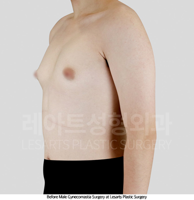
Before starting a detailed consultation,
a visual examination revealed clear protrusion
in both sides of the patient’s chest.
The most prominent bulging was observed
around the nipple and areola,
which had been the patient’s main concern.
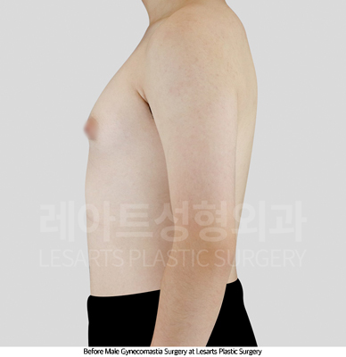
On palpation, a firm tissue mass
was felt beneath the nipple,
suggesting glandular hypertrophy.
To confirm this, an ultrasound examination
was performed, which showed deep and wide overgrowth
of glandular tissue beneath both nipples.
Based on these findings,
the patient was diagnosed with true gynecomastia,
corresponding to Simon Grade 2A.
To treat the condition effectively,
surgical removal of the glandular tissue
was determined to be necessary.
1.Gland Excision
Through an areolar incision,
the enlarged glandular tissue—the root cause of gynecomastia—
was completely removed.
2.Chest Liposuction
Liposuction was performed around the glands
and along the lateral chest area
to remove residual fat and refine the contour,
creating a smooth, natural chest shape.
3.Adhesion Prevention & Tissue Repositioning
To prevent nipple depression or skin adhesion
after gland removal,
tissue repositioning was done to ensure
a balanced and even chest contour.
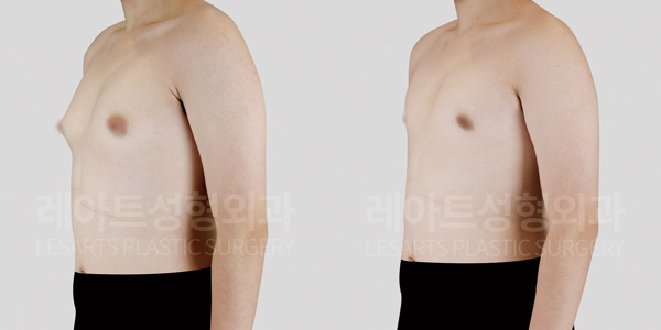
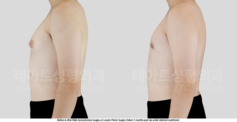
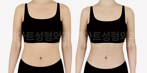
At 2 months post gynecomastia surgery,
we reviewed and compared
the patient’s chest contour improvements
to preoperative photographs.
Before surgery, the areola and nipple appeared
significantly enlarged and protruding,
with the surrounding chest forming a rounded bulge.
The lower chest and nipple area were especially pointed,
creating a noticeably raised contour.
Because of this, attention was often drawn
to the chest line, and when wearing thin clothing,
the protrusion became more visible,
causing discomfort and self-consciousness.
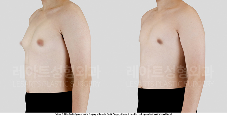
At 2 months after gynecomastia surgery,
the glandular tissue was removed,
and chest liposuction was also performed.
As a result, the overall chest volume
was naturally reduced,
and the bulging around the nipple and areola
disappeared, leaving a smooth, stable contour.
In particular, the sharp projection of the chest
was corrected, resulting in a flat, masculine shape
with a well-balanced and natural appearance.
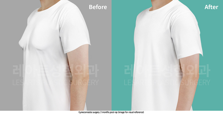
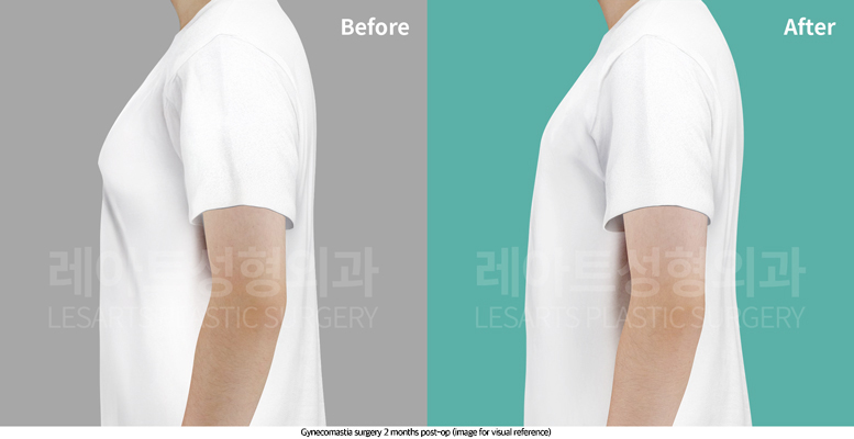
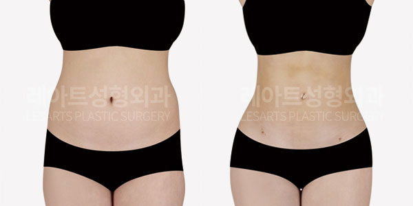
After gynecomastia surgery,
the chest contour under a T-shirt now appears
firm and smooth, resembling the results
of consistent strength training.
Before surgery, the chest bulge and protruding nipples
were easily visible through thin clothing,
often causing self-consciousness and discomfort.
Following gland excision and lateral chest liposuction,
the chest size was effectively reduced,
and the nipple area naturally flattened,
eliminating those aesthetic concerns.
The patient reported that not only the chest shape improved,
but also the intermittent pain and pressure
around the nipples that had persisted for years
had completely disappeared after surgery.
He expressed high satisfaction with
both the cosmetic and functional improvements.
As seen in this case, true gynecomastia—
caused by excessive glandular tissue growth—
often involves not only visible protrusion or enlargement,
but also physical discomfort such as pain or tenderness.
Therefore, if similar symptoms are present,
it is important to consult with a qualified specialist
for an accurate diagnosis and appropriate surgical treatment plan,
rather than delaying medical evaluation.
If you are experiencing similar discomfort or concerns,
consider discussing gynecomastia treatment options
with your medical team sooner rather than later.
#Gynecomastia
#MaleBreastReduction
#TrueGynecomastia
#ChestLiposuction
#GlandExcision
#MenChestSurgery
#BeforeAndAfter
#LesartsPlasticSurgery
#KoreanPlasticSurgery
#BestLiposuctionClinicInKorea